Neuroanatomy Self Portrait
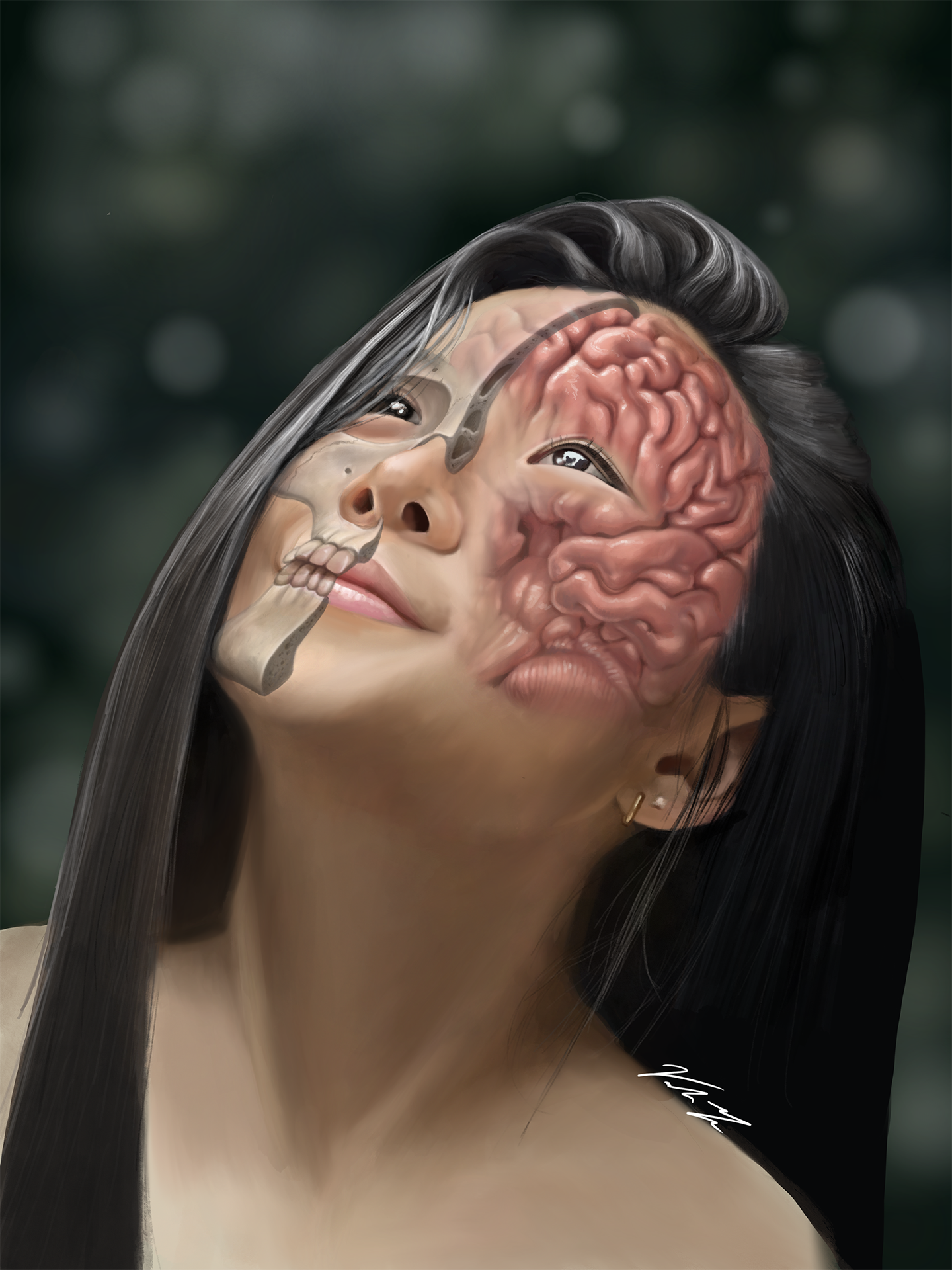
The purpose of this illustratio is to create an anatomically correct editorial
piece with a focus on external neuroanatomy. One of the challenges with this assignment was positioning
the brain correctly within the skull.
To learn how this piece came to be, and how I overcame that challenge, continue reading.
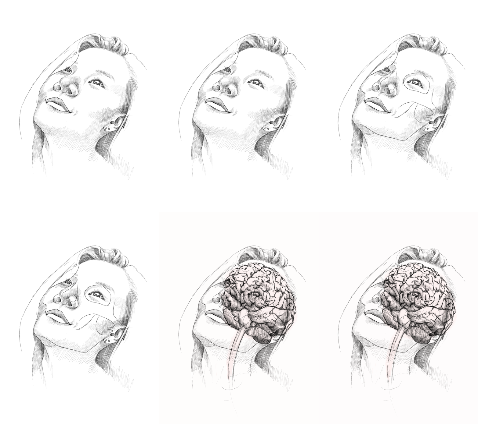
Sketching out the General Postions
Using photo reference, a self portrait was quickly sketched out. Then referencing textbooks, online 3D models, real human skulls, and plastic brain models, the general position of both the skull and brain is mapped out.

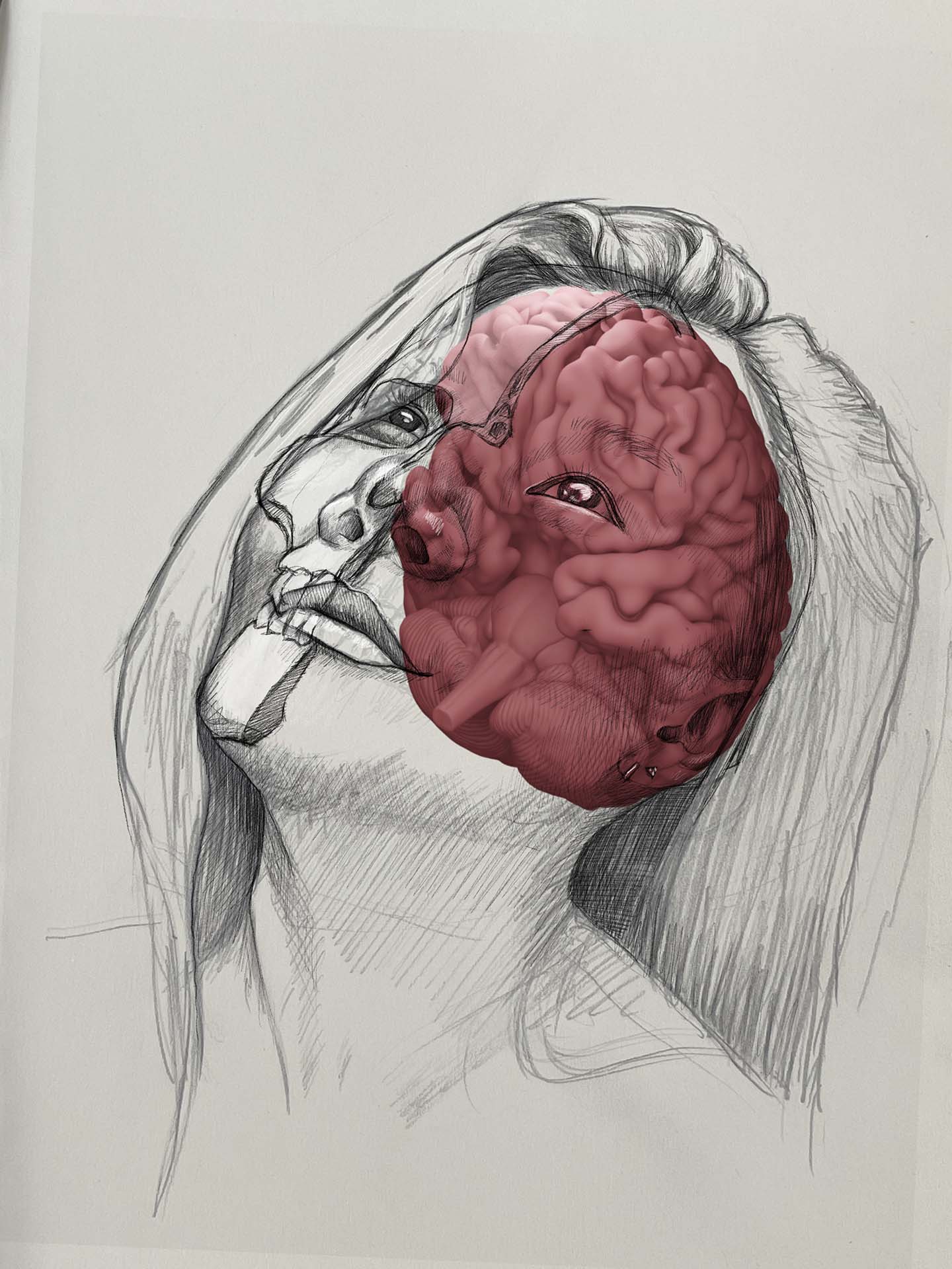
3D Maquette
Using a 3D brain model, I was able to position it to mirror the same angle that my head is tilted at and then rendered it out. I then overlayed the brain model on top of the portrait sketch as so that the 3D model can be used as a maquette for where the brain should be drawn. By checking the oribitomeatal plane aligns with the bottom of the temporal lobe of hte brain, I can see that the brain is positioned in the correct orientation and position.

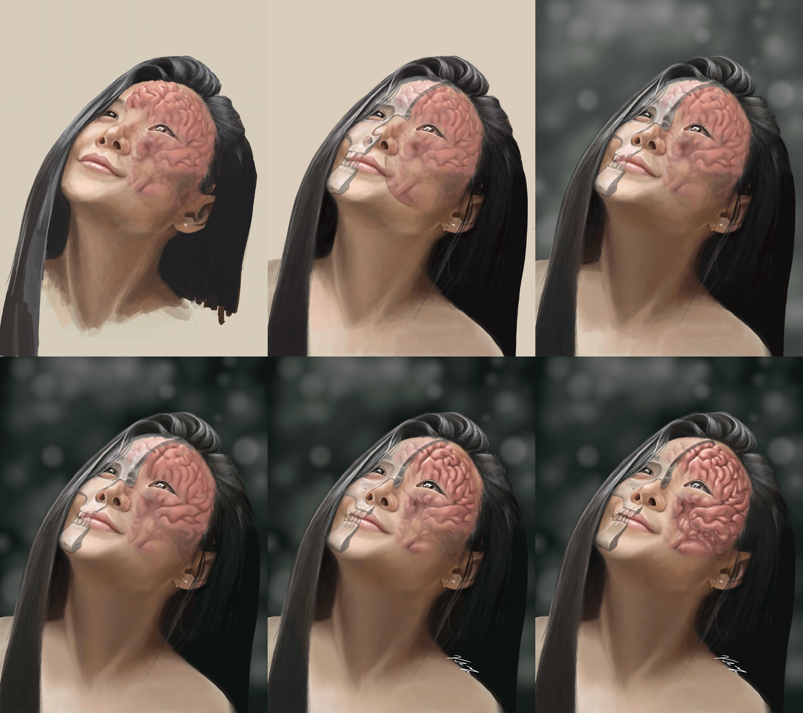
Final Rendering
After researching and checking that the anatomical position of both the skull and brain are correct, the piece is rendered via digital painting with special care taken to the different textures found in the skull and brain. For the brain coloration, fresh lamb's brain was acquired and observed.
